
When presenting summary cancer statistics, sometimes the oral cavity and pharynx are linked together (see Chapter 20: Oral Cavity and Pharynx in the SEER Cancer Statistics Review (CSR), 1975‒2015).
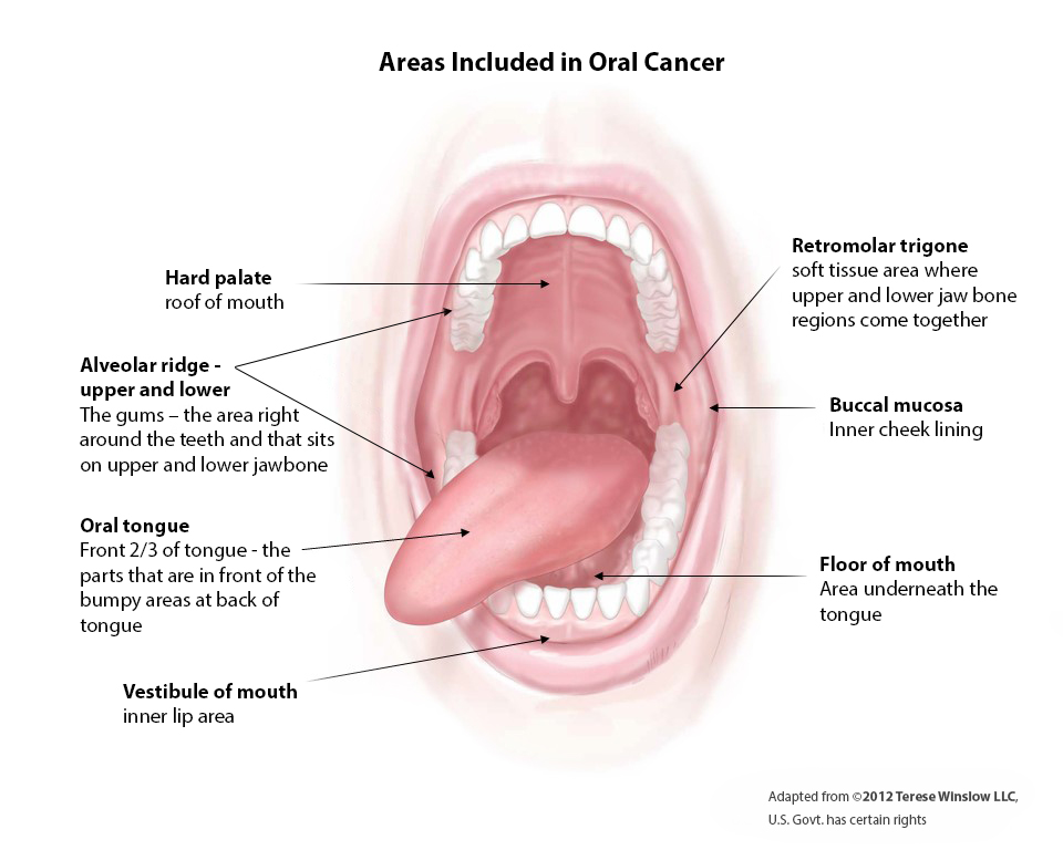
- The oral cavity includes the hard palate, gums, mobile tongue, retromolar trigone, cheek, inner lip, and floor of the mouth (see figure).
- The oral cavity is separate from the:
- oropharynx, which includes the tongue base, tonsils, soft palate and the associated throat mucosa; and
- nasopharynx, which includes the adenoid lymphoid tissue, the mucosa of the back of the nose, and the nasal side of the soft palate; and
- hypopharynx, which includes the low, lateral and posterior walls of the throat (below where the naked eye can see when looking into a person’s open mouth).
The oropharynx, nasopharynx, and hypopharynx—which collectively make up the pharynx—are not included in this calculator. The prognosis of cancers located in the head and neck varies a lot by site, so it is important to separate them into their logical groupings. Reasons for the different prognosis include:
- the different risk factors for cancer at each site, the largest three of which are tobacco, alcohol, and human papillomavirus (HPV); For example, oral cavity cancers are strongly associated with alcohol and tobacco use, but not HPV. In contrast, oropharynx cancer is strongly associated with HPV;
- natural anatomic boundaries, which tend to allow some cancers to spread more easily than others; For example, the prognosis of larynx cancer is much better than hypopharynx cancer, even though the two locations are quite near each other.